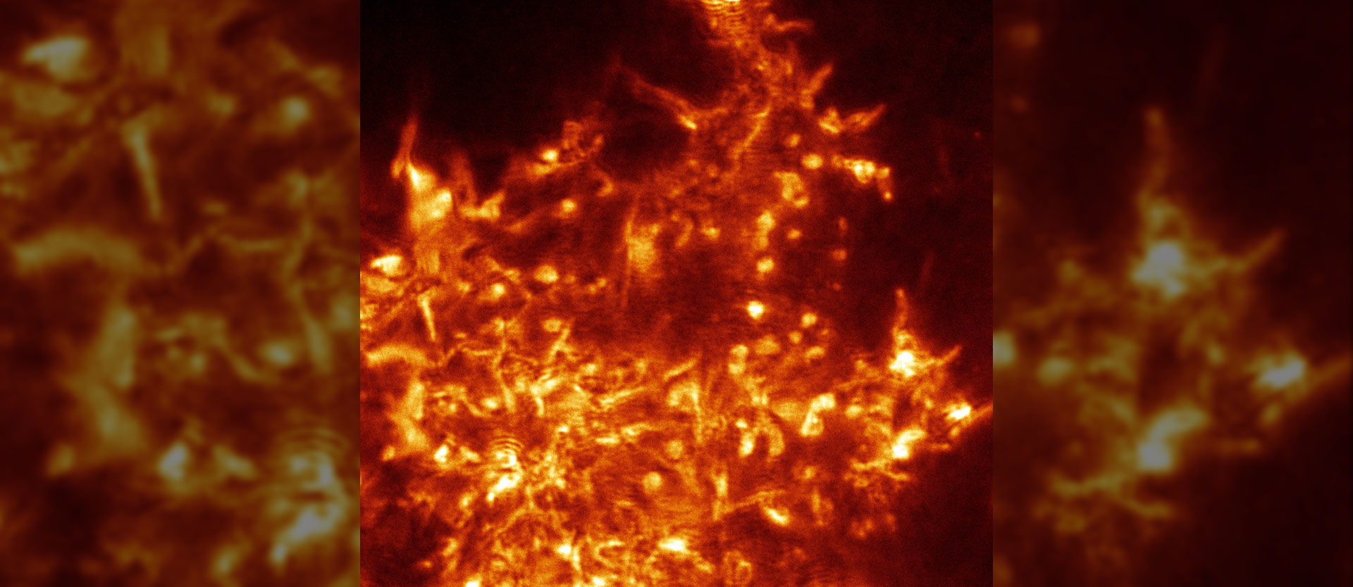
Cell dynamics under blue laser light
Using a new laser method, a team led by Prof. Dr. Alexander Rohrbach from the Laboratory of Bio- and Nano-Photonics at the University of Freiburg has found a way to make the smallest cell structures visible in detail without fluorescence.

The technique used is called "rotating coherent scattering" (ROCS) and is based on a blue, rapidly rotating laser beam. "We use several physical phenomena known from everyday life," Rohrbach says. Small objects such as molecules, viruses or cell structures scatter blue light most strongly, as is known, for example, from air molecules in the atmosphere and what we perceive as blue sky. "Small objects scatter and direct about ten times more blue than red light particles to the camera, thereby transmitting valuable information," Rohrbach explains.
In addition, ROCS directs a blue laser at a very oblique angle to the biological objects because this significantly increases contrast and resolution. This, too, is familiar from everyday life: Dirt is easier to see when a wine glass is held at an angle against the light. Third, the scientists illuminate the object successively from all sides with the oblique laser beam, because only one direction of illumination alone would produce many artifacts.
100 images per second of living cells
The Freiburg physicists and engineers rotate the oblique laser beam around the object one hundred times per second, generating 100 images per second. "So in ten minutes, we already have 60,000 images of living cells, which are much more dynamic than previously thought," Rohrbach says. However, such dynamic analyses of just one minute of image material require enormous computing power from computers. Here, various computer algorithms and analysis methods had yet to be developed in order to interpret the data correctly.
Together with his colleague Dr. Felix Jünger and in cooperation with various research groups in Freiburg, Rohrbach was able to demonstrate the microscope's performance on various cell systems: "It was not our goal to produce beautiful images or movies of the unexpectedly high dynamics of cells - we wanted to gain new biological insights." For example, it was possible to observe for the first time how stimulated mast cells open small pores in just a few milliseconds to shoot out spherical granules with inexplicably high force and speed. The granules contain the messenger substance histamine, which can later lead to allergic reactions.
Virus-like particles observed
In further experiments, the researchers observed virus-like particles dancing around the fissured surface of phagocytes at a ludicrous pace, only to find a point of attachment to the cell after a few attempts. These observations served as preliminary experiments to studies currently underway on the attachment behavior of coronavirus.
In addition, ROCS technology has been used to study scar formation in cardiac muscle injury. Fibroblasts, cells of scar tissue, form 100-nanometer-thin tubes called nanotubes that are 1,000 times thinner than a hair. Using the new technique, the researchers discovered that these tubes vibrate thermally on tiny scales, but that this motion subsides over time.
In addition, the scientists saw how the filopodia - the "fingers" of phagocytes - scan their surroundings for prey in a complex quivering motion, and how their cytoskeleton changes at a previously unknown rate.
Original publication:
Jünger, F., Ruh, D., Strobel, D., Michiels, R., Huber, D., Brandel, A., Madl, J., Gavrilov, A., Mihlan, M., Daller, C. C., Rog-Zielinska, E.A., Römer, W., Lämmermann, T.,Rohrbach, A. (2022):
100 Hz ROCS microscopy correlated with fluorescence reveals cellular dynamics on different spatiotemporal scales.
Nature Communications 13(1): 1758.
doi.org/10.1038/s41467-022-29091-0


