Deep inside the mouse brain
Researchers at the Duke Center for In Vivo Microscopy have created incredibly sharp images of a mouse brain using a combination of magnetic resonance imaging and light-sheet microscopy.
 t is an unusual play of colors that opens up before our eyes. It provides us with completely new and extremely detailed insights into the brain of a mouse. We have the mix of greatly improved magnetic resonance imaging (MRI) and light sheet microscopy to thank for these views. The combined imaging technique was developed by scientists at the Duke Center for In Vivo Microscopy and with the collaboration of colleagues at the University of Tennessee Health Science Center, the University of Pennsylvania, the University of Pittsburgh and Indiana University.
Magnetic resonance imaging just celebrated its 50th birthday. The imaging technique is able to visualize soft tissue that is difficult to visualize with X-rays. MRI images now resolve well enough to detect a brain tumor, but they still need to be much sharper to reveal microscopic details in the brain.
Duke researchers have now taken an important step toward that goal. They have created a scan of the mouse brain that is much sharper than a typical human clinical MRI. A single so-called voxel in the new images - think of it as a cubic pixel - measures just five microns. That's 64 million times smaller than a clinical MRI voxel.
The refined MRI thus offers an entirely new way to visualize the neural connectivity of the entire brain at record-breaking resolution. As a result, it is now possible to better observe how the brain changes with age, diet, or even neurodegenerative diseases such as Alzheimer's, for example.
"This is something that's really going to take us forward. We can start to look at neurodegenerative diseases in a very different way," says G. Allan Johnson, Ph.D., the study's lead author.
The improved imaging is made possible by an extremely powerful 9.4 Tesla magnet. Most clinical MRIs use a magnet of 1.5 to 3 tesla. This is joined by a special set of gradient coils that are 100 times more powerful than those in a clinical MRI. And last but not least was a high-powered computer connected to it, with a computing capacity of nearly 800 laptops.
After Johnson and his team scanned the brain, they sent the tissue for imaging using light sheet microscopy. This additional technique allows them to label specific groups of cells, such as dopamine-producing cells, to monitor the progression of Parkinson's disease. The scientists then overlaid the light sheet microscope images with the original MRI scan, which provides a view of cells and circuits throughout the brain.
With this mix of data, researchers can now explore the microscopic mysteries of the brain in ways that were not possible before. For example, one series of MRI images shows how the connections change as the mice age, but also how certain regions, such as the subiculum involved in memory, change more than the rest of the mice's thinking organ.
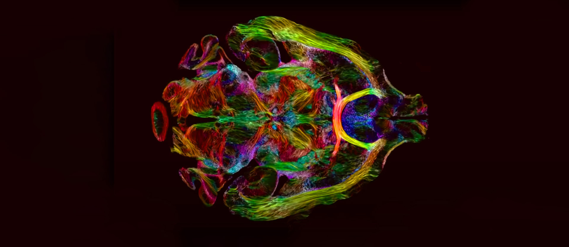
Another series of images shows a series of rainbow-colored brain connections that illustrate the deterioration of neural networks in a mouse brain with Alzheimer's disease.
Original publication:
G. Allan Johnson, Yuqi Tian, David G. Ashbrook, Gary P. Cofer, James J. Cook, James C. Gee, Adam Hall, Kathryn Hornburg, Yi Qi, Fang-Cheng Yeh, Nian Wang, Leonard E. White, Robert W. Williams.
Merged Magnetic Resonance And Light Sheet Microscopy Of The Whole Mouse Brain
Proceedings of the National Academy of Sciences, 17. April 2023. DOI: 10.1073/pnas.2218617120
Welcome to Photonworld – a platform for knowledge, interesting facts and science on the topic of light. Photonworld aims to provide a basic understanding of the physics of light and its countless phenomena. From medicine, to art and physics: all topics are covered.
Photonworld originated as an initiative of the excellence cluster the Munich-Centre for Advanced Photonics (MAP) and the Laboratory for Attosecond Physics (LAP) at the Max Planck Institute of Quantum Optics (MPQ).


The initiator of the project is Prof. Dr. Ferenc Krausz, Chair of Experimental Physics at the LMU and Director of the MPQ.
Photonworld’s Student Zone reports directly out of the PhotonLab, which is supported by the Munich Center for Quantum Science and Technology (MCQST) and the DFG Research Unit FOR 2783. At the PhotonLab, schoolteachers and students can find up-to-date information about experiments and goings-on at the lab, and plan their own visit to the laboratory.